Medical imaging systems make it possible for health care professionals to see processes within the body so they can diagnose, monitor or treat medical problems. It’s a growing industry — one market research company is forecasting growth from $30.2 billion in 2013 to $49 billion in 2020.
Oakland University researcher Jing Tang, Ph.D., ABSNM, and her small team in the Biomedical Imaging Laboratory in the School of Engineering and Computer Science, are part of the imaging revolution that is improving diagnosis and treatment.
Dr. Tang, assistant professor in the department and director of the imaging laboratory, leads research designed to improve medical image reconstruction, evaluation, and analysis techniques. More specifically, Dr. Tang, two graduate students and a postdoctoral researcher are working on algorithms that, when incorporated into equipment software, will improve the images generated by certain imaging systems.
Dr. Tang’s current research, funded by a National Science Foundation CAREER Award, involves data from the emerging and cutting-edge hybrid PET/MRI (positron emission tomography/magnetic resonance imaging) biomedical imaging system. PET imaging, she says, measures functional information in the body, while MRI imaging provides anatomical information with better soft tissue contrast than a computer tomography (CT) — scan.
The hybrid imaging system is relatively new, having received Food and Drug Administration approval in 2011. The technology is used primarily for oncology, neurology and cardiology applications.
“Whether the two systems are used sequentially or simultaneously, the hybrid PET/MRI system gives physicians more detailed and thorough information than a single system scan,” Dr. Tang says. “Most prefer simultaneous scans, but even with both options in place, there are more technical difficulties to overcome,” she adds.
Collaborating with OUWB School of Medicine
Dr. Tang’s research into how to improve the images generated by the hybrid imaging systems involves close collaboration with Oakland University William Beaumont School of Medicine physicians.
“They are our real-world connections,” Dr. Tang says. “They tell us what kind of image problems they would like us to solve and we develop algorithms to address them. For example, a doctor might say there are artifacts in certain areas of the images, and ask us to find a solution to that problem.”
The work involves determining how to overcome obstacles involved with forming the image as well as how to make the most of the integrated data generated by both imaging methods — PET and MRI.
“The images generated by the data collected in the scans are already very good,” Dr. Tang notes, “but there’s room for improvement. For example, organ movement and signal loss when traveling through the body both have an impact on the resulting image that physicians study. How can we overcome those issues?”
The goal, she says, is to use modern techniques to extract more information from the data to create even higher quality images. The work could, ultimately, contribute to providing patients with personalized medicine.
“The better the image, the better able physicians will be to see what’s going on and decide on the best, most targeted treatment,” she says.
[visibility type=”hidden-phone”]
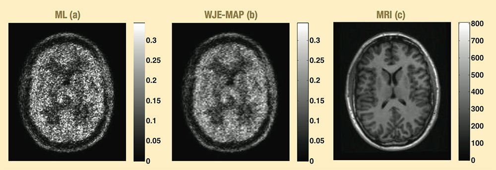
The brain PET image from a patient radiotracer (11C-DPA-713) study reconstructed using (a) the conventional reconstruction method and (b) the newly developed reconstruction method incorporating the anatomical information from (c) the corresponding MR image. The new method demonstrates its potential in clinical quantitative PET imaging. 11C-DPA-713 is a promising radiotracer for evaluating translocator protein (TSPO) binding with PET. TSPO can serve as a marker of neuro-inflammation.
Sharing developments
The research results will eventually get translated into code that will be incorporated into medical imaging equipment software.
More immediately, the team’s research results are shared in medical specialty conference papers and presentations. For example, team members made two presentations at the Society of Nuclear Medicine and Molecular Imaging annual meeting in June, including “Anatomy-assisted direct 4D parametric image reconstruction for dynamic cardiac PET imaging.”
Dr. Tang also received funding through the National Science Foundation’s Broadening Participation Research Initiation Grants in Engineering to reach and engage historically under-represented minority students. Working with undergraduate students, she created and presented to Detroit-area high school students the “I See You” workshop on biomedical imaging.
“We want to interest underrepresented students, including young women, in engineering careers in general but also in biomedical engineering and biomedical imaging careers specifically,”
Dr. Tang says. “We’ve recently been able to collaborate with the School of Engineering and the School of Medicine, so we have a greater variety of presenters and presentations.”
Providing better patient care
As Dr. Tang continues her research to improve the images generated by hybrid PET/MRI technology, she remains focused on her end goal: Better care for patients.
“For me, it’s not about publishing high-profile papers or moving up through the ranks in academia, although that’s all good. My ultimate goal is to contribute to the well-being of people throughout the world,” Dr. Tang says.
In fact, it is why she chose medical imaging over offers in other fields that included the oil industry.
“Improving health care is what’s in my heart,” she says. “My lab’s contribution right now is tiny, but we’re doing things that will help.”
Source: Post by Sandra Beckwith, Oakland University